
In rodents, stroke is most frequently modeled by transient occlusion of the middle cerebral artery (tMCAO), in which a coated monofilament is introduced into the vasculature to occlude the artery for the period intended by the researcher 1, 5. Rodents are the preferred animal model in nervous system research, due to their similarity to humans-in anatomy, biochemistry, and physiology-, and low maintenance and processing in the laboratory 4. Despite important pre-clinical findings using stroke animal models, the lack of translation into clinical practice is pointed as the major factor that delays the implementation of new therapies 3. The only treatment available focuses on reperfusion of the brain areas affected, via endovascular thrombectomy or blood clot-busting with tPA (tissue plasminogen activator) 2. Ischemic stroke is the leading cause of mortality and disability and the second leading cause of dementia in western countries 1. This methodology proves to be a breakthrough in the field, by providing a precise and detailed assessment of stroke outcomes in preclinical animal studies.

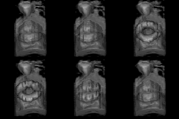
Of note, whole brain 3D reconstructions allow brain atlas co-registration, to identify the affected brain areas, and correlate them with functional impairment. In the transient-ischemic-attack (TIA) mouse model, we can quantify striatal myelinated fibers degeneration. We used the intraluminal transient MCAO (Middle Cerebral Artery Occlusion) mouse stroke model to identify and quantify ischemic lesion and edema, and segment core and penumbra regions at different time points after ischemia, by manual and automatic methods. Here, we successfully describe the application of brain contrasting agents (Osmium tetroxide and inorganic iodine) for high-resolution micro-CT imaging for fine location and quantification of ischemic lesion and edema in mouse preclinical stroke models. In the last decade, high-resolution micro-CT for 3D sample analysis turned into a simple, fast, and cheaper solution.

Until recently, the analysis of brain lesions was performed using two techniques: (1) histological methods, such as TTC (Triphenyltetrazolium chloride), a time-consuming and inaccurate process or (2) MRI imaging, a faster, 3D imaging method, that comes at a high cost. Characterization of brain infarct lesions in rodent models of stroke is crucial to assess stroke pathophysiology and therapy outcome.


 0 kommentar(er)
